shandilya 02
18yr old male with bilateral weakness and edema of lower limbs
Case: 18 year old with chief complaints of bilateral weakness and edema of lower limbs
- I've been given this case to solve in an attempt to understand the topic of "patient clinical data analysis" to develop my competency in reading and comprehending clinical data including history, clinical findings, investigations and come up with a diagnosis and treatment plan.
You can find the entire real patient clinical problem in this link here made by our interns
https://hitesh116.blogspot.com/2020/05/12may-2020-elog-medicine-intern.html?m=1
https://srianugna.blogspot.com/2020/05/hello-everyone.html
Following is my analysis of this patient's problem:
Chief complaints:
The problems in order of priority I found are
Weakness of both lower limbs since 20 days
Bilateral edema in lower limbs.
History of presenting illness in the order of priority
1-B/L weakness in lower limbs
Weakness initially started in the proximal region 2 years back .
Onset: insidious in onset ,
Progression:progressed gradually to distal region
Associated features
-difficulty in wearing and holding chappals
-difficulty in combing hair , buttoning and unbuttoning shirt.
---B/L calf hypertrophy present
Possible diagnosis:
From above chief complaints we can rule out certain factors and come into final conclusion.
Weakness may be caused by any one of the following related to blood vessels,muscle or nervous system.
If Artery related
As in case of peripheral artery disease and varicose veins there will be pain,leg skin atrophy,and claudication,telengectasia. As there are no signs of this artery are ruled out.
If suspecting nerves related?
May be it is related to nerves because from the history there is history of difficulty in holding chappals and difficulty in buttoning and unbuttoning shirt.
To understand the nerve related issue A thorough CNS EXamination is done and following are results.
CNS-EXAMINATION
patient is conscious, coherent, coperative
patient well oriented to time, place and person
higher mental functions= normal
Cranial nerves- intact
Motor system-
tone - normal
power - 4-/5 in both lower limbs
reflexes absent in both lower limbs
sensory system-normal
No meningeal signs
No cerebellar signs
1- ANATOMICAL LOCATION OF THE PROBLEM
From the above CNS examination it is observed that there might be lesion in LMN pathway.
LMN LESION can be particularly divided into
1)neurogenic(anterior horn cell and peripheral nerves)
2) neuromuscular junction
3) muscle
If suspecting neurogenic cause:
Here nerve conduction test is done which is normal so we can rule out neurogenic cause
If suspecting Neuromuscular junction cause
Electromyography is done which is normal and thus neuromuscular junction disorders like myesthenia gravis and lamborten Eaton syndrome are ruled out .
Finally we left with muscular cause.
Muscle biopsy should be done and examination of creatine kinase levels must be done.
Creatine levels -increased

Muscle biopsy report(quadriceps femoris)- suggesting no evidence of POLYMYOSIS and may be suggestive of dystrophy.


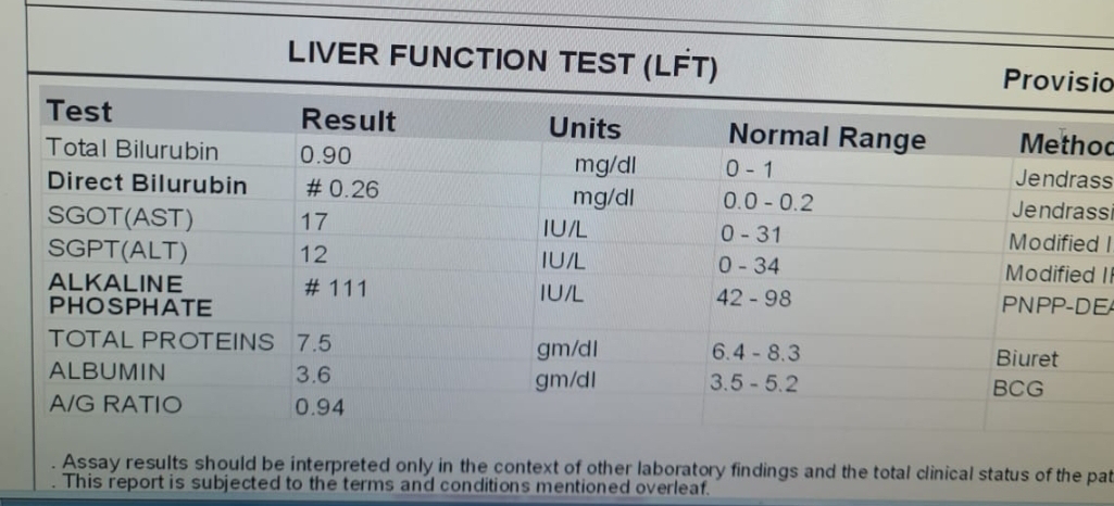
Other investigations
1.CBP -normocytic normochromic with leucocytosis
2.serology
3.RFT-creatinine and uric acid levels are slightly elevated
4.ECG- normal
5.CUE- pus cells seen
are done and these reports are within normal range.
2-ETIOPATHOGENESIS OF ABNORMALITY PRESENT IN MUSCLE (MYOPATHIES)
We have to rule out each and every myopathies now as follows
1)INFLAMMATORY MYOPATHIES
2} METABOLIC MYOPATHIES
3)DRUG INDUCED MYOPATHIES
4)ENDOCRINE MYOPATHIES
5)MUSCULAR DYSTROPHY
1)INFLAMMATORY myopathies can be ruled out as there are no antibodies produced and no signs of inflammation are seen
2) Metabolic myopathies can also be ruled out as we can see there are no signs of glycogen storage disordersetc
3)Drug induced myopathies can also be ruled out
as there is no relavent history of drug intake from patient.
4)endocrine myopathies can also be ruled out as there are no thyroid Abnormalities and thyroid function is normal
5) It is most likely the muscular dystrophy cause
Because proximal muscle weakness of lower extremities occur first and muscle biopsy report shows fat cells and necrotising and regenerative muscle fibres are seen suggesting dystrophy.
NOW WE HAVE TO DIAGNOSE WHICH TYPE OF MUSCULAR DYSTROPHY??
There are 3 most common types of dystrophies seen
1)myotonic dystrophy
2)Duchene's muscular dystrophy
3)Becker's muscular dystrophy
and the other one which is unlikely limb girdle muscular dystrophy
We have to rule out one by one and come to final diagnosis
1) myotonic dystrophy
This can be ruled out easily as it starts with facial muscle Weakness and sparing proximal muscles mostly.
2)Duchene's muscular dystrophy
It can also be ruled out easily as it presents early in 3 to 5 years of age and patients shows Gower's manuever while standing up.
They usually die within 16-18 yes with severe pulmonary infections
Joint contractures and scoliosis are other features seen.
3)Becker's muscular dystrophy
Here Muscle wasting resembles Duchene's
Here proximal muscle weakness of lower extremities occur first
Onset 5-15 years of age
Dystrophin muscle protein is absent
Calf muscles shows hypertrophy due to accumulation of fat cells and necrotic tissue.
Therapeutic options
Reference
https://www.google.com/amp/s/www.hopkinsmedicine.org/health/conditions-and-diseases/becker-muscular-dystrophy%3famp=true